Patient Education
Technology & InnovationIntraoral Cameras: Seeing What Your Dentist Sees
A small camera captures detailed images of every tooth surface. Learn how intraoral photography improves diagnosis, communication, and insurance documentation.
- Technology & Innovation
- Patient Guide
- Dental Tips
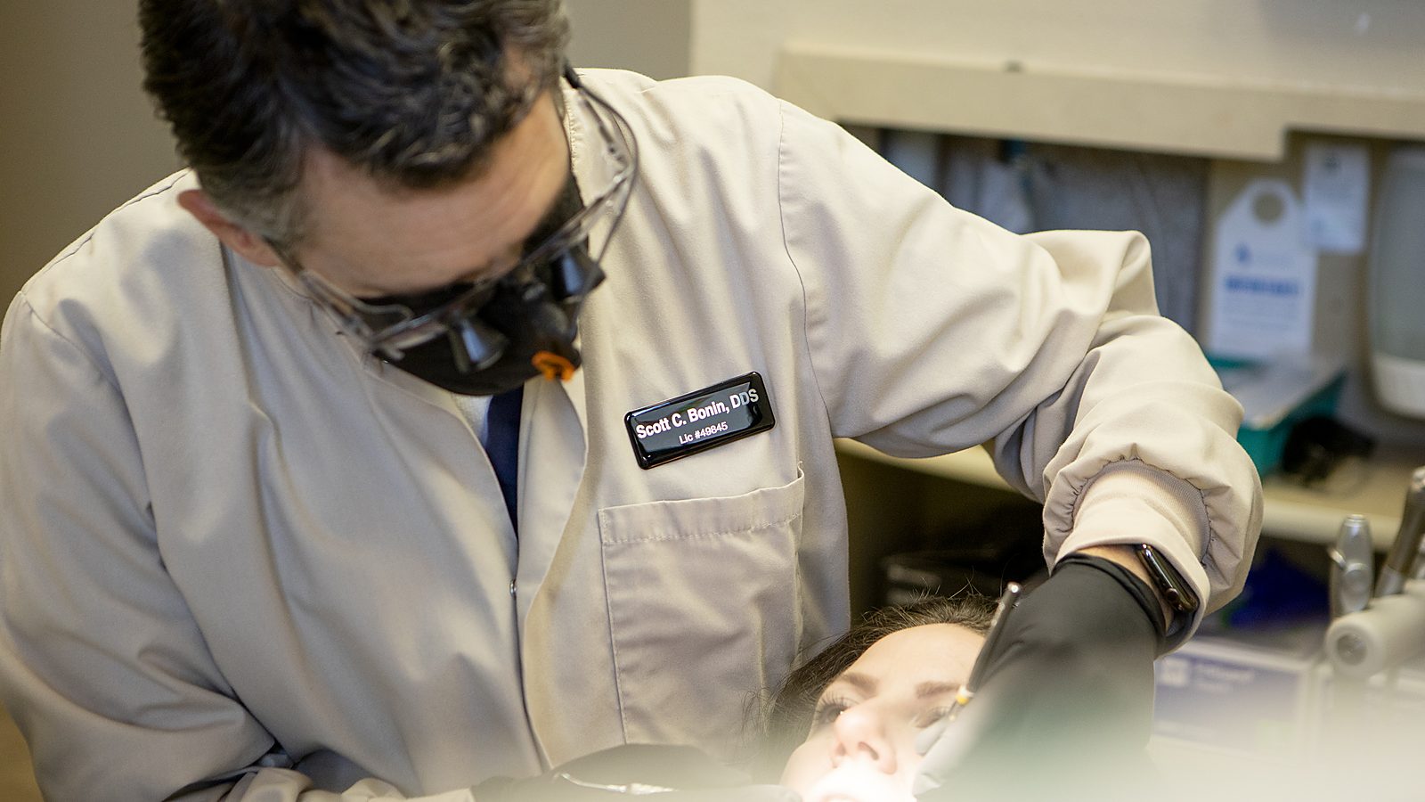
When your dentist looks in your mouth, they see details that are hard to describe to you sitting in the chair. A small crack, a subtle color change, an area of decay, and these clinical observations don’t translate easily into words. Intraoral cameras solve this communication gap by showing you exactly what Dr. Bonin sees. A tiny camera captures high-resolution images of your teeth and gums from every angle. These images improve diagnosis, enhance communication, and create documentation that strengthens insurance claims.
How Intraoral Cameras Work
An intraoral camera is a small handheld device with a camera and light source at the tip. When placed against a tooth or in an area of your mouth, it captures detailed photographs. Unlike a smartphone camera that can’t focus on small objects up close, intraoral cameras use specialized optics to photograph teeth and gum tissue at magnifications where small details become visible.
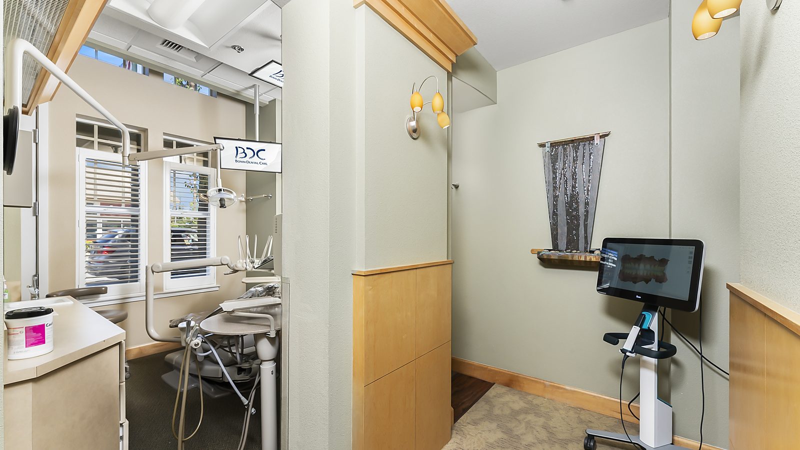
The images are displayed on a chairside monitor so you can see them in real-time. Dr. Bonin can point to areas on the screen, explaining what he’s seeing and what treatment is recommended. You’re not just hearing his explanation; you’re seeing the actual tissue.
Most modern intraoral cameras are digital, storing images directly to your treatment record. These images become part of your permanent dental documentation, creating a visual record of your oral health over time.
Improved Diagnosis and Documentation
Intraoral photography improves diagnosis by creating clear documentation of conditions. A cavity visible on X-ray can be photographed from multiple angles, documenting exact location and extent. This documentation is invaluable if the cavity requires insurance pre-authorization or if a claim is disputed.
Gum disease documentation is particularly valuable. Inflammation, recession, pocket depth, and tissue color changes are all more obvious in photographs than in words alone. Insurance companies reviewing claims for gum disease treatment can see the actual condition in photographs, making approval more likely.
Crowns and other restorations can be photographed before treatment, creating a baseline for comparison. If issues arise years later, you have baseline images showing the tooth’s original condition. This documentation strengthens arguments about what happened and what should be retreated.
Enhanced Patient Communication
Many patients delay or deny treatment because they don’t understand the clinical problem. Seeing a cavity in an intraoral photograph makes the problem concrete and real. You can see the brown discoloration, understand why treatment is needed, and commit to it.
The same applies to other conditions. Seeing calculus buildup on X-rays is one thing; seeing the actual yellow calculus deposits and inflamed gum tissue in an intraoral photograph is far more persuasive. Patients who see photographic evidence of disease are more motivated to pursue treatment and follow prevention recommendations.
Dr. Bonin uses intraoral photographs during consultations to show you exactly what he’s recommending treatment for. This visual evidence makes recommendations easier to understand and accept.
Insurance Documentation and Claims
Insurance companies increasingly require photographic documentation for claims. When you claim a filling is needed, the insurer wants documentation of decay. When you claim gum disease treatment is needed, the insurer wants evidence of disease. Intraoral photographs provide this evidence directly.
Predetermination requests submitted with intraoral photographs are more likely to be approved because the insurance company can see exactly what treatment is being recommended and why. The photographs remove guesswork and make claims processing faster.
If a claim is denied and you appeal, intraoral photographs strengthen your appeal. You can show the insurer that treatment was indeed medically necessary because the photographic evidence is irrefutable.
Comparing Treatment Over Time
Intraoral photographs create a visual record of your oral health across years. Comparing photographs from your first visit to current images shows whether your oral health is improving, stable, or declining. This visual comparison is motivating; you can literally see the improvement from better home care or the decline if disease is progressing.
For patients with gum disease, comparing photographs across years shows whether scaling and improved home care are working. Seeing gum recession halted or tissue color improved reinforces that your efforts are succeeding.
For cosmetic cases, before and after photographs document treatment results. If you’re considering teeth whitening or other cosmetic work, before photographs show your baseline, and after photographs show the improvement.
Limitations and Considerations
Intraoral photography requires time. Taking comprehensive photographs of all surfaces adds minutes to your appointment. Some patients find having a camera in their mouth uncomfortable, though most quickly adapt.
Image quality depends on camera quality and operator skill. Reflections, angles, and lighting can affect image clarity. A skilled operator can generally capture clear images, but some cameras produce better images than others.
Privacy is a consideration. Intraoral photographs are stored in your treatment record and shared with dental labs or specialists if treatment requires it. While images are protected as medical records, the concept of having photographs of your mouth in a system might make some patients uncomfortable. This is why practices should explain their photography policy and obtain consent before photographing.
Intraoral photographs are supplemental to other diagnostic tools. They don’t replace X-rays, clinical examination, or other imaging. They augment these tools by providing visual documentation.
Cosmetic Applications
Intraoral photography is particularly useful for cosmetic dentistry. Before pursuing cosmetic dentistry like veneers, teeth whitening, or smile makeovers, photographs document your baseline. You can discuss specific changes you’d like to see and have them clearly documented.
During digital smile design, intraoral photographs are used as the baseline for digital simulations. You see what your smile might look like after treatment before any procedures are done.
Intraoral Photography at Bonin Dental Care
Dr. Bonin uses intraoral photography during your exam and treatment planning. You’ll see the images on the chairside monitor, and Dr. Bonin will explain what they show. The photographs become part of your treatment record and support insurance claims and documentation.
If you have concerns about photography or privacy, let us know. We respect your preferences about photographic documentation and can discuss how images are stored and used.
Technology Enhancing Communication
Intraoral photography represents how technology can improve communication between dentist and patient. Instead of abstract descriptions of problems, you see concrete visual evidence. This clarity helps you make informed decisions about treatment and understand why recommendations are made.
Understanding modern diagnostic tools helps you appreciate their role in improving your care. When Dr. Bonin recommends treatment, you’re not just trusting his words; you’re seeing photographic evidence of why treatment is needed. This visual clarity enhances your confidence in recommended treatment.
Schedule a consultation at Bonin Dental Care in Windsor, California to experience how intraoral photography enhances your dental care and communication with Dr. Bonin.
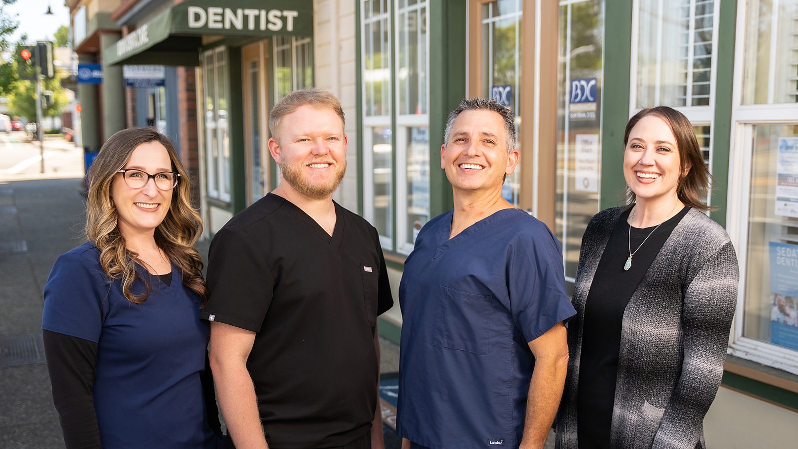
Written by
Dr. Scott Bonin, DDSGeneral and cosmetic dentist at Bonin Dental Care in Windsor, California. USC School of Dentistry graduate, Navy veteran, and member of the American Dental Association, California Dental Association, and American Academy of Cosmetic Dentistry. Over 24 years of clinical experience serving Sonoma County families.
View full credentialsClinical note: This article is for educational purposes and does not replace a professional examination. Every patient's situation is unique. If you have questions about your specific dental health, please schedule an appointment or call (707) 838-1400.
Related Services
Explore the treatments behind this topic
Ready to talk with Dr. Bonin about what you just read? Here are the procedures at Bonin Dental Care most closely connected to this article. Each page explains how we do the work, what to expect, and how to get started.
-
General & Preventive
General Dentistry
Comprehensive exams, professional assessments, and preventive strategies designed to catch problems early and keep your smile healthy.
Learn about this service -
General & Preventive
Dental Cleanings
Professional cleanings remove tartar and buildup that home care cannot reach, preventing decay and gum disease.
Learn about this service
Ready to book your visit with Dr. Bonin?
New patients welcome. Call (707) 838-1400 or request an appointment online.
Keep Reading
More from our library
-
Electronic Health Records in Dentistry: How They Improve Your Care
Digital records enable better coordination, faster referrals, and comprehensive treatment histories. Learn how EHR systems benefit patients directly.
-
Teledentistry: When a Virtual Visit Makes Sense
Virtual consultations work for follow-ups, second opinions, and triage. Learn what teledentistry can and cannot evaluate remotely.
-
How Modern Dental Materials Have Changed in the Last Decade
Zirconia, lithium disilicate, bioactive cements, and nano-composites have transformed durability and aesthetics. A look at what is different and why it matters.