Patient Education
Technology & InnovationCone Beam CT Scans in Dentistry: What They Show That X-Rays Can't
CBCT provides 3D views of bone, nerve canals, sinuses, and root anatomy. Learn when this imaging is recommended and how it improves treatment planning.
- Technology & Innovation
- Dental Implants
- Patient Guide
Traditional X-rays show a two-dimensional flat image of your teeth and jawbone. For many situations, this is sufficient. But for complex cases, X-rays miss important information that only three-dimensional imaging reveals. Cone Beam Computed Tomography (CBCT), sometimes called a dental CT scan, captures your entire jaw in 3D detail, showing bone structure, nerve pathways, sinus anatomy, and tooth roots in ways traditional X-rays cannot. This technology has transformed treatment planning for complex cases, particularly dental implants and surgical procedures.
How CBCT Works
A CBCT scanner rotates around your head, capturing hundreds of individual X-ray images from different angles. Computer software reconstructs these images into a complete 3D model of your jaw, teeth, bone, and surrounding structures. You stand still inside the machine for about 20-30 seconds while the scan captures. Radiation exposure is higher than a single X-ray but much lower than traditional medical CT scans.
The resulting 3D image can be viewed from any angle, allowing Dr. Bonin to see your anatomy in complete detail. He can measure bone volume, identify nerve locations, assess sinus positions, and evaluate tooth root anatomy in three dimensions instead of the flat, distorted view a traditional X-ray provides.
What CBCT Shows That X-Rays Don’t
Traditional X-rays are two-dimensional projections. They show flat images that overlap and distort, making precise three-dimensional assessment difficult. CBCT eliminates this limitation, showing exact bone heights, widths, and densities. This precision is crucial for guided implant surgery.
CBCT clearly identifies the inferior alveolar nerve, the major nerve running through the lower jaw. Knowing its exact position before implant surgery prevents nerve damage. Traditional X-rays show the nerve canal’s approximate location, but CBCT shows it with precision allowing guided implant placement that avoids the nerve entirely.
CBCT also shows sinus anatomy. If you need implants in the upper back jaw (posterior maxilla), knowing sinus position and size is critical. CBCT determines whether implants can fit above the sinus or if sinus augmentation (adding bone) is needed first. Traditional X-rays can’t make these determinations reliably.
Root anatomy is shown in detail with CBCT. For complex root canals or tooth extractions, CBCT reveals root positions and configurations. Curved roots, multiple roots, and internal resorption are all clearly visible. This information helps Dr. Bonin plan treatment and warn you about potential complications before they occur.
CBCT also assesses bone density and quality. Some bone is dense and strong; other bone is softer and resorbs easily. CBCT shows this variation, allowing implant planning that accounts for bone quality. Implants in soft bone need different approaches than implants in dense bone.
When CBCT is Recommended
CBCT is recommended when implant planning requires precise bone assessment. Before placing dental implants, Dr. Bonin needs to know bone height, width, and density. CBCT provides this information definitively. Any multi-implant case or complex single implant definitely warrants CBCT.
Surgical cases benefit from CBCT. Impacted wisdom teeth, complicated extractions, and bone augmentation procedures all benefit from 3D planning. A patient might know they have a wisdom tooth that needs extraction, but not realize how its roots relate to the nerve. CBCT reveals this, allowing proper surgical planning.
Endodontic cases (root canals) sometimes benefit from CBCT if the case is complex or the diagnosis unclear. Root resorption, curved anatomy, and missed canals are all more clearly identified on CBCT. For straightforward root canals, traditional imaging suffices, but complex cases benefit from 3D information.
Orthodontic cases involving implants or unusual bone anatomy sometimes warrant CBCT. Oral surgeons planning bone grafts use CBCT to assess donor and recipient sites. Any surgery involving critical anatomical structures benefits from CBCT planning.
CBCT and Radiation Safety
CBCT exposes you to radiation, so Dr. Bonin doesn’t recommend it without clinical justification. However, the radiation dose is quite low, typically less than 30-100 microsieverts depending on scanner type. For comparison, you receive about 100 microsieverts annually just from background radiation in the environment.
The risk of radiation-induced cancer from a single CBCT is extremely low, much lower than many common medical risks we accept without hesitation. The benefit of precise surgical planning that prevents nerve damage or implant failure typically far outweighs the minimal radiation risk.
Dr. Bonin orders CBCT only when he believes the diagnostic information is important enough to justify the radiation exposure. He won’t recommend CBCT for routine diagnostic purposes where traditional X-rays suffice. But for surgical cases where precision matters, CBCT is standard of care.
CBCT and Guided Implant Surgery
CBCT is central to guided implant surgery. The 3D scan is used with special software to plan implant position down to the millimeter. The computer even generates a surgical guide, a 3D-printed template that directs the implant drill at the precise angle and depth planned in software. Guided implant surgery using CBCT and 3D-printed guides dramatically improves precision and reduces complications compared to traditional freehand implant placement.
Patients receiving guided implant surgery have more predictable outcomes, faster healing (because surgical trauma is minimized), and better long-term implant success. The combination of CBCT imaging and guided surgery represents state-of-the-art implant dentistry.
Cost and Insurance
CBCT adds cost to treatment. The scan itself costs money, and not all insurance plans cover it. Some plans require documentation of medical necessity before they’ll pay. At Bonin Dental Care, we submit insurance authorization requests and work with your plan to obtain coverage. If your plan doesn’t cover CBCT, we discuss the cost with you and help you decide whether the diagnostic benefit justifies the out-of-pocket expense.
For implant cases, CBCT is almost always worthwhile. The cost is small relative to implant treatment costs, and the diagnostic information directly impacts implant success. For cases where traditional imaging might suffice, we discuss with you whether CBCT would improve diagnosis and justify the cost.
The Digital Workflow
CBCT scans integrate seamlessly with CAD/CAM dentistry and digital treatment planning. The 3D scan data can be combined with digital impressions to create a complete virtual model of your anatomy and planned treatment. This allows precise prediction of treatment outcomes before treatment begins.
For complex cases, Dr. Bonin can use CBCT data with planning software to show you exactly where implants will be placed and what the final result will look like. You see the treatment plan in 3D before any drilling begins.
CBCT at Bonin Dental Care
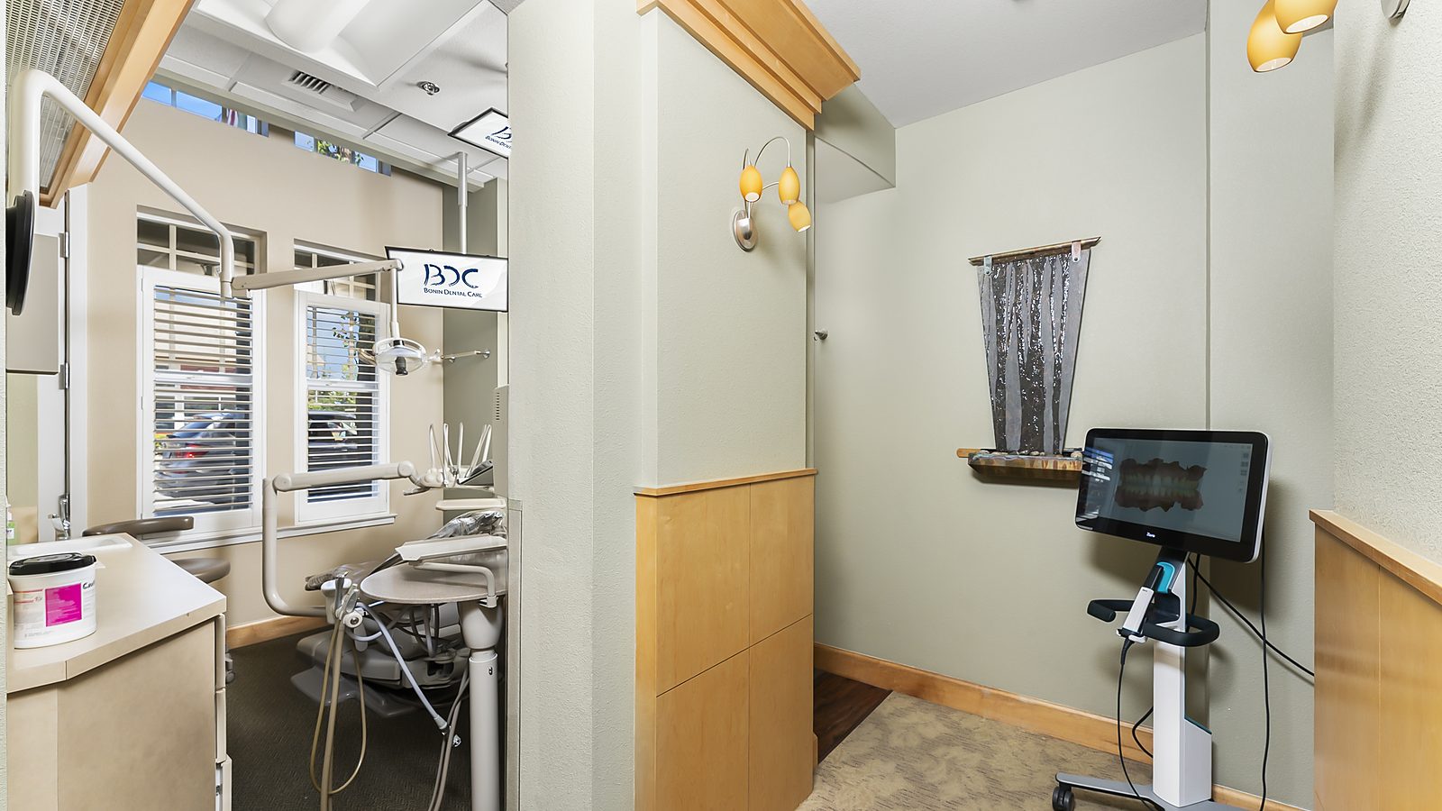
Bonin Dental Care has on-site CBCT capability, meaning Dr. Bonin can capture scans conveniently during your appointment. This integrates seamlessly into your treatment workflow. Scans are captured, analyzed immediately, and used for treatment planning without delay or additional appointments.
If you’re facing complex dental treatment like implant surgery or complicated extractions, Dr. Bonin may recommend CBCT. We’ll discuss the cost, benefits, and radiation considerations, then use the scan to create a precise treatment plan that gives you the best possible outcome.
Understanding modern dental technology like CBCT helps you appreciate why certain diagnostic tools are recommended. When Dr. Bonin suggests advanced imaging, he’s recommending it because the diagnostic information will improve your care. Schedule a consultation at Bonin Dental Care in Windsor, California to discuss your treatment options and how advanced technology can optimize your outcomes.
Written by
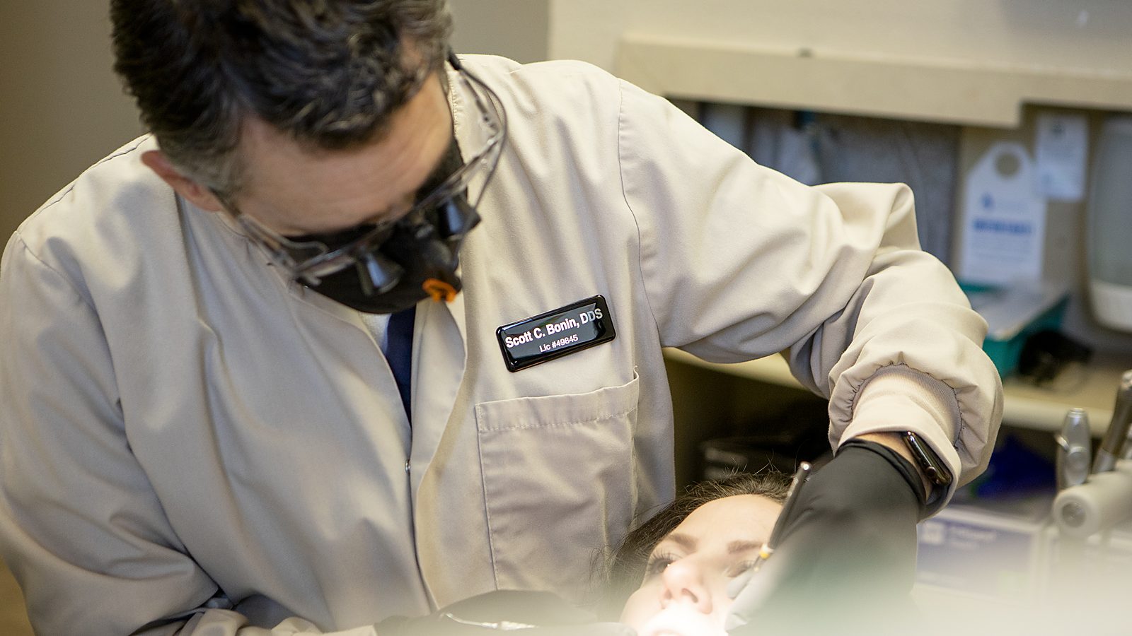
Dr. Scott Bonin, DDSGeneral and cosmetic dentist at Bonin Dental Care in Windsor, California. USC School of Dentistry graduate, Navy veteran, and member of the American Dental Association, California Dental Association, and American Academy of Cosmetic Dentistry. Over 24 years of clinical experience serving Sonoma County families.
View full credentialsClinical note: This article is for educational purposes and does not replace a professional examination. Every patient's situation is unique. If you have questions about your specific dental health, please schedule an appointment or call (707) 838-1400.
Related Services
Explore the treatments behind this topic
Ready to talk with Dr. Bonin about what you just read? Here are the procedures at Bonin Dental Care most closely connected to this article. Each page explains how we do the work, what to expect, and how to get started.
-
Restorative
Dental Implant Restoration (Surgical Care Coordinated with Specialists)
Dr. Bonin coordinates the surgical placement of your dental implant with a trusted oral surgeon or periodontist, then designs and seats the custom crown that finishes the case at our Windsor office.
Learn about this service -
Restorative
Full-Arch Dental Implants (All-on-4 / All-on-X, Coordinated with Specialists)
Full-arch restoration with All-on-4 or All-on-X implants. The surgical phase is performed by a trusted oral surgeon Dr. Bonin coordinates with, and Dr. Bonin designs and seats the final prosthesis at our Windsor office.
Learn about this service -
Restorative
Implant-Supported Dentures (Coordinated with Surgical Specialists)
Implant-supported dentures snap onto two to four implants and stay secure all day. Dr. Bonin coordinates the surgical phase with a trusted oral surgeon and designs the custom denture and attachments at our Windsor office.
Learn about this service
Ready to book your visit with Dr. Bonin?
New patients welcome. Call (707) 838-1400 or request an appointment online.
Keep Reading
More from our library
-
Electronic Health Records in Dentistry: How They Improve Your Care
Digital records enable better coordination, faster referrals, and comprehensive treatment histories. Learn how EHR systems benefit patients directly.
-
Teledentistry: When a Virtual Visit Makes Sense
Virtual consultations work for follow-ups, second opinions, and triage. Learn what teledentistry can and cannot evaluate remotely.
-
Guided Implant Surgery: Precision Through Technology
Computer-guided surgery uses CT scans and 3D-printed guides to place implants with sub-millimeter accuracy. Learn the process and its advantages.